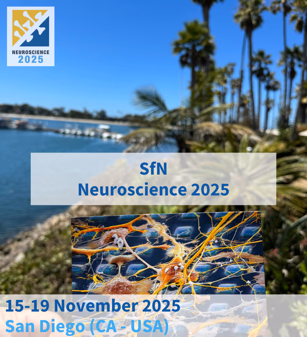
Neuroscience 2025 (SfN)
Date & Time
Location
Tags
The Society for Neuroscience (SfN) Annual Meeting is the world’s leading event dedicated to advancing the understanding of the brain and nervous system.
Each year, tens of thousands of researchers, clinicians, and industry professionals gather to share groundbreaking discoveries and explore the latest innovations in neuroscience.
Recognized as the largest and most influential conference in the field, Neuroscience 2025 will feature five days of inspiring talks, symposia, poster sessions, and hands-on exhibits at the San Diego Convention Center.
We are excited to be back in this year’s meeting: visit our booth to discover our latest technologies, meet our team, and see what’s new!
We will also be presenting posters, hosting a special satellite event, and sharing a few surprises at our booth throughout the week.
If you would like to attend but need support, we offer a Travel Grant for SfN 2025: check it out here.
Meet the MxW Team

Dr. Carlos Sanchez Priego

Dr. Francesca Puppo

Dr. Laura D'Ignazio

Dr. Marian Hruska-Plochan

Dr. Marie Obien

Miguel Veloso O'Donell

Dr. Silvia Oldani

Dr. Tom Dufor

Dr. Urs Frey

Dr. Xiaohan Xue
%2520Li.avif)
Dr. Zhuoliang (Ed) Li
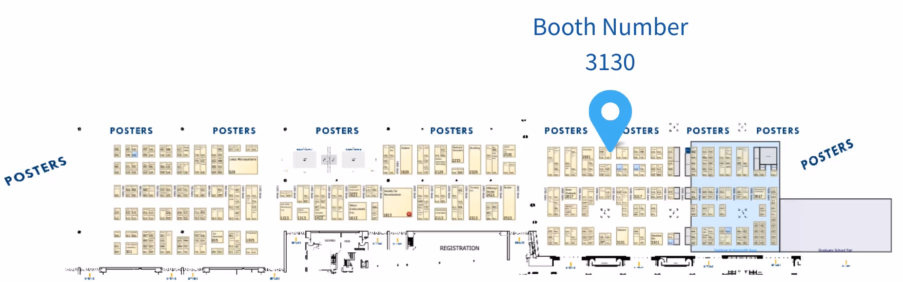
MxW Booth
Find us at booth #3130: come meet us in person and explore what’s new!
Poster Presentations
Dr. Marian Hruska-Plochan
Sunday, November 16, 2025, 1:00-5:00 PM
Abstract
Human neural models of amyotrophic lateral sclerosis (ALS) and frontotemporal dementia (FTD) are essential for advancing research of these fatal neurodegenerative disorders. The advent of iPSC-derived neural systems, from neuronal monocultures to brain and spinal cord organoids, has enabled discoveries that were previously impossible due to the limited availability of live patient CNS tissue, the human-specific RNA-binding of TDP-43, cross-species barriers, and the general inadequacy of animal models of these diseases.
To fully leverage these complex human-derived neural networks, understanding their functional properties, and how these are altered in disease, is a prerequisite. This need is further underscored by recent NIH and FDA policy shifts that prioritize human-based research models, such as organoids and tissue-on-chip platforms, over traditional animal models. As biological research enters this new era, deciphering the functional dynamics of these advanced systems is more important than ever.
In this study we applied our next-generation High-Density Microelectrode Arrays (HD-MEAs), which enable real-time, label-free, and non-invasive electrophysiological recordings with unmatched spatial resolution, allowing to fully characterize and exploit the complexity of these in vitro neural cultures. The MaxTwo multiwell HD-MEA platform, featuring 26,400 electrodes per well, captures activity across entire networks, individual neurons, and subcellular compartments, including axonal arbors.
Using this system, we profiled disease-specific activity in ALS (TDP-43 M337V, Q331K) and FTD (GRN KO) models. The platform’s ultra-dense coverage and industry-leading signal-to-noise ratio (SNR) enabled consistent detection of disease phenotypes with minimal sample replicates. ALS cultures exhibited reduced spontaneous and network activity, while GRN KO neurons displayed pronounced hyperactivity and irregular oscillations, phenotypes that would likely be missed by low-density MEAs due to signal averaging and sparse spatial coverage.
To probe subcellular alterations, we applied our revolutionary AxonTracking assay to extract metrics such as conduction velocity, latency, axonal length, and branching by tracing action potential propagation. GRN KO neurons exhibited significantly shorter, less-branched axons, suggesting that their hyperactivity may represent a compensatory response to structural deficits.
Our HD-MEA platforms combine high-resolution electrophysiology with automated tools for data visualization, batch analysis, and rapid report generation, enabling robust, reproducible phenotyping and screening. This scalable system is ideally suited for acute and longitudinal studies in ALS and FTD research and therapeutic development.
Biography
Dr. Zhuoliang (Ed) Li
Tuesday, November 18, 2025, 1:00-5:00 PM
Abstract
Advances in Microelectrode Array (MEA) technology for in-vitro electrophysiological recordings have made it possible to study neuronal networks across multiple scales, from subcellular properties to network-level dynamics. These devices are essential for exploring the phenotypes of neurological disorders and accelerating drug discovery, offering unique insights into the behaviour of neuronal networks. Key factors such as electrode density, spacing, and size significantly impact signal quality, noise, and sensitivity. To exhaustively characterize neuronal networks, MEAs must combine single-cell and subcellular resolution with high-throughput capabilities, maintaining sensitivity to small extracellular action potentials to capture the full range of network activity. In this study, the MaxOne and MaxTwo high-density (HD) MEA systems (MaxWell Biosystems, Switzerland) were utilized to record activity from induced pluripotent stem cell-derived neurons. These systems, with 26,400 electrodes per well, demonstrated the benefits of increased statistical power in longitudinal data collection. HD-MEA recordings were compared to simulated low-density recordings, where adjacent electrodes on HD-MEAs were clustered to mimic larger, lower-density electrodes. Additionally, the AxonTracking Assay, an automated tool for analysing individual axonal arbours from multiple neurons simultaneously, was used to evaluate axonal structures and network functionality in the recorded cultures. Results showed that higher electrode density and smaller electrode size enhanced sensitivity, allowing for the detection of smaller spikes and capturing the complete spectrum of network dynamics. The high-resolution analysis of network activity, combined with subcellular insights from the AxonTracking Assay, offers a robust platform for drug screening and disease modelling.
Biography
Ed is a well-traveled and trained neuroscientist, having received his Bachelors of Science at the University of Toronto, Masters in Neuroscience at the University of Munich, and PhD in Neurobiology at the University of Basel. Ed has demonstrated his wide range of expertise and experience in in vitro and in vivo electrophysiology, calcium imaging, circuit mapping, behavior, as well as neuropharmacology through his publications on the circuitry underlying depression and anxiety. Since 2022, Ed has been a member of MaxWell Biosystems, where he is currently the Product Manager for the MaxOne Single-Well High Density Microelectrode Array System.
When he is not shaping the future of electrophysiology, Ed is all about bold moves, whether it is skiing, exploring new countries, or strategizing imaginative and unconventional ways to beat opponents in trading card games.
Do you want to learn more?
Book a one-to-one call with one of our experts to discuss how MaxWell Biosystems HD-MEA platforms can empower your research!