Pharmacology & Toxicology
Every Cell has a Story to Tell.
Let’s Discover Yours
Pharmacology and toxicology studies with in vitro biological systems allow researchers to evaluate how compounds modulate neuronal function and assess potential safety risks in a controlled, human-relevant context.
Researchers can monitor activity patterns and network dynamics to assess the efficacy, potency and adverse effects of small molecules, biologics or environmental agents on neuronal cultures, organoids or brain slices. These readouts provide essential information on dose-response relationships, therapeutic windows, and compound-specific mechanisms of action, offering translational biomarkers and functional endpoints relevant in clinical trials. When combined with complementary approaches like imaging, transcriptomics, and structural analysis, in vitro pharmacology and toxicology provide a comprehensive view of how compounds influence neuronal health and function from single cells to complex networks.
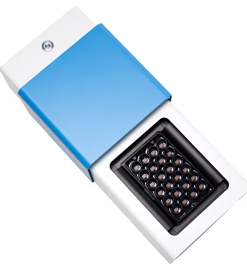
MaxWell Biosystems’ HD-MEA platforms offer unmatched resolution and throughput for pharmacological and toxicological screening. With label-free, high-content recordings from thousands of electrodes across multi-well formats, researchers can detect subtle functional changes, monitor compound effects over time, and compare responses across conditions or cell lines. This enables robust dose-response profiling, safety assessment, and mechanism-of-action studies, all within a scalable, automation-compatible platform designed for reproducibility and translational relevance.
Consistent drug responses with high reproducibility
Pharmacological and toxicological responses can vary across cell types, doses, and replicates. Reproducible functional readouts across wells and timepoints help separate true compound effects from technical noise or variability caused by limited signal coverage, enabling confident, data-driven decisions.
Track compound effects on axonal function
Many compounds influence axonal conduction and growth. High-resolution axon tracking reveals changes in conduction velocity and morphology, helping researchers detect toxicity-related impairments or therapeutic rescue effects on axonal function.
Detect subtle compound effects early
Compound effects may first appear as slight changes in spiking, network dynamics, or excitability. Sensitive detection allows researchers to capture both low-amplitude signals and subtle shifts in activity, enabling early identification of therapeutic action or emerging toxicity.
Scale up screening with confidence
Screening compounds across doses, timepoints, or cell lines requires consistent, high-throughput analysis. Our automation-ready HD-MEA platforms support robust data acquisition and processing, enabling efficient evaluation of drug efficacy, mechanism-of-action, or safety profiles.
Relevant biological models:



Case studies

Dose-dependent response to synaptic blockers
This study used the MaxTwo HD-MEA Multi-Well System to profile the functional response of Elixirgen Scientific (Ricoh Biosciences) Quick-Neuron™ human iPSC-derived excitatory neurons to increasing doses of the synaptic blockers NBQX (AMPA receptor antagonist) and D-AP5 (NMDA receptor antagonist). High-content HD-MEA recordings revealed a rapid and dose-dependent suppression of neuronal firing and network bursting with NBQX, while D-AP5 produced more modest decreases. Recovery of activity after drug washout confirmed assay stability, showcasing robust, reproducible pharmacological evaluation.
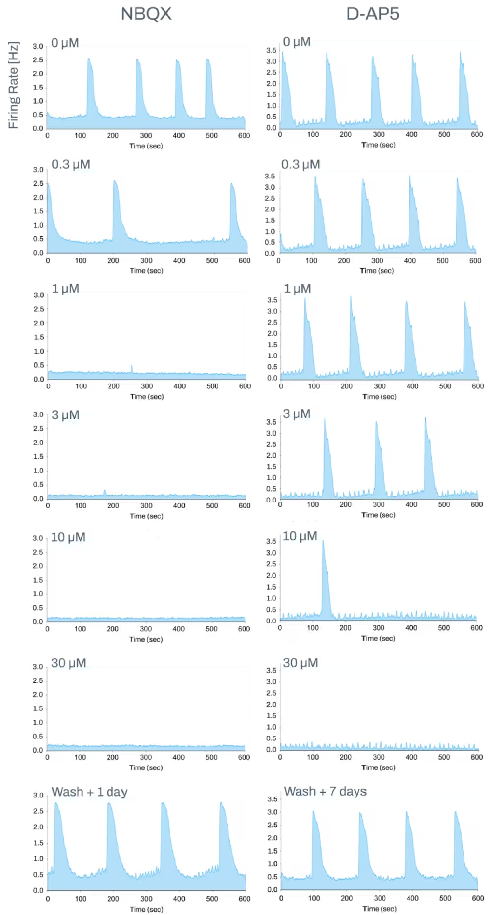

Dose-Dependent Effects of NBQX and D-AP5 on Firing Rates in Ricoh Biosciences Quick-Neuron™ Human Excitatory Neurons
Representative network activity plots show suppression of spontaneous network bursts with increasing concentrations of AMPA/Kainate receptor antagonist NBQX (left column) and NMDA receptor antagonist D-AP5 (right column). Washout panels demonstrate recovery of network activity, confirming reversibility and robustness of pharma assay measurements using the MaxTwo HD-MEA system.
Data adapted from the Application Note in collaboration with Elixirgen Scientific (Ricoh Biosciences).
Scalable pharmacological validation of different neuroactive compounds on 3D InSight™ Human Neural Microtissues
The HD-MEA MaxTwo 24-Well Plate enables scalable, label-free functional profiling of InSphero 3D InSight™ Human Neural Microtissues with high consistency and reproducibility across wells. ActivityScan and Network Assays capture robust spontaneous activity, synchronized bursting, and stable network phenotypes, providing a powerful baseline for compound testing.
Pharmacological modulation with standard neuroactive compounds such as kainic acid, 4-AP, isoguvacine, and TTX revealed clear, differential network responses, ranging from altered bursting dynamics to complete activity suppression. These results highlight how HD-MEA technology unlocks high-throughput pharmacological validation in complex 3D neuronal models, supporting reliable compound-response screening with network-level insight.
.avif)
Scalable HD-MEA profiling reveals consistent network activity and differential compound responses in 3D neuronal microtissues.
Top: Representative ActivityScan Assay firing-rate maps from four independent 3D InSight™ Human Neural Microtissues demonstrate consistent functional activity. Confocal imaging shows microtissue morphology, while raw unfiltered traces from three microtissues highlight robust high-amplitude action potentials across time scales. Scale bars: 100 µm.
Bottom: Representative baseline and post-treatment (20 mins after) recordings demonstrating compound-specific network modulation. Kainic acid, 4-AP, isoguvacine, and TTX produced distinct electrophysiological signatures, highlighting the sensitivity of HD-MEA recordings for pharmacological validation and compound-response profiling in 3D neuronal models.
Data obtained in collaboration with InSphero AG.
Longitudinal serotonin treatment promotes network maturation in cerebellar organoids
The MaxOne HD-MEA System was utilized for monitoring long-term pharmacological effects in human iPSC-derived cerebellar organoid models. Cerebellar organoids were chronically treated with serotonin (5-HT), a compound known to support Purkinje cell maturation. Longitudinal high-resolution HD-MEA recordings revealed that serotonin-treated cerebellar organoids developed robust, synchronized network bursting activity, a key functional indicator of advanced synaptic maturation. Our HD-MEA technology allows sensitive, non-invasive tracking of functional changes in organoids over time, supporting detailed analysis of compound effects on neuronal network development.
.avif)
Effect of chronic serotonin treatment on network activity in human iPSC-derived cerebellar organoids
Top: ActivityScan Assay firing rate maps and Network Assay network activity plots reveal sparse, unsynchronized firing in untreated organoids at DIV 56.
Bottom: Cerebellar organoids chronically treated with serotonin (5-HT) display pronounced, synchronized network bursting and elevated firing rates at DIV 56, indicating enhanced synaptic network maturation.
Data obtained in collaboration with Ana Maria Gomes from the Stem Cell Engineering Research Group (SCERG), Lisbon.
Selected Resources
Application of a high-density microelectrode array assay using a 3D human iPSC-derived brain microphysiological system model for in vitro neurotoxicity screening of environmental compounds

Analgesic effect of Botulinum toxin in neuropathic pain is sodium channel independent

P97/VCP ATPase inhibitors can rescue p97 mutation-linked motor neuron degeneration


Dr. Marcus Kaji
The improved signal-to-noise ratio with high density is a huge advantage for drug screening. We can use lower drug concentrations to see effects across the entire chip, whereas low-density setups tend to show only an average effect, requiring higher doses. High density lets us detect the susceptibility of lower-firing neurons to compound silencing, which adds a new layer of insight. It gives us higher pharmacological resolution, which is invaluable for characterizing drug responses as well as potency and efficacy.
.avif)
Dr. Maria Sundberg
“To identify the molecular pathways that are causing the disease phenotypes in these neurons we have studied their network functions on MaxWell Biosystems HD-MEA, gene expression, profiles with RNA sequencing, and synaptic marker expression profiles using immunocytochemical assays. The multi-well platform allows us to record from several wells/conditions at the same time. This is helpful when comparing the network function between control and patient cell populations.”