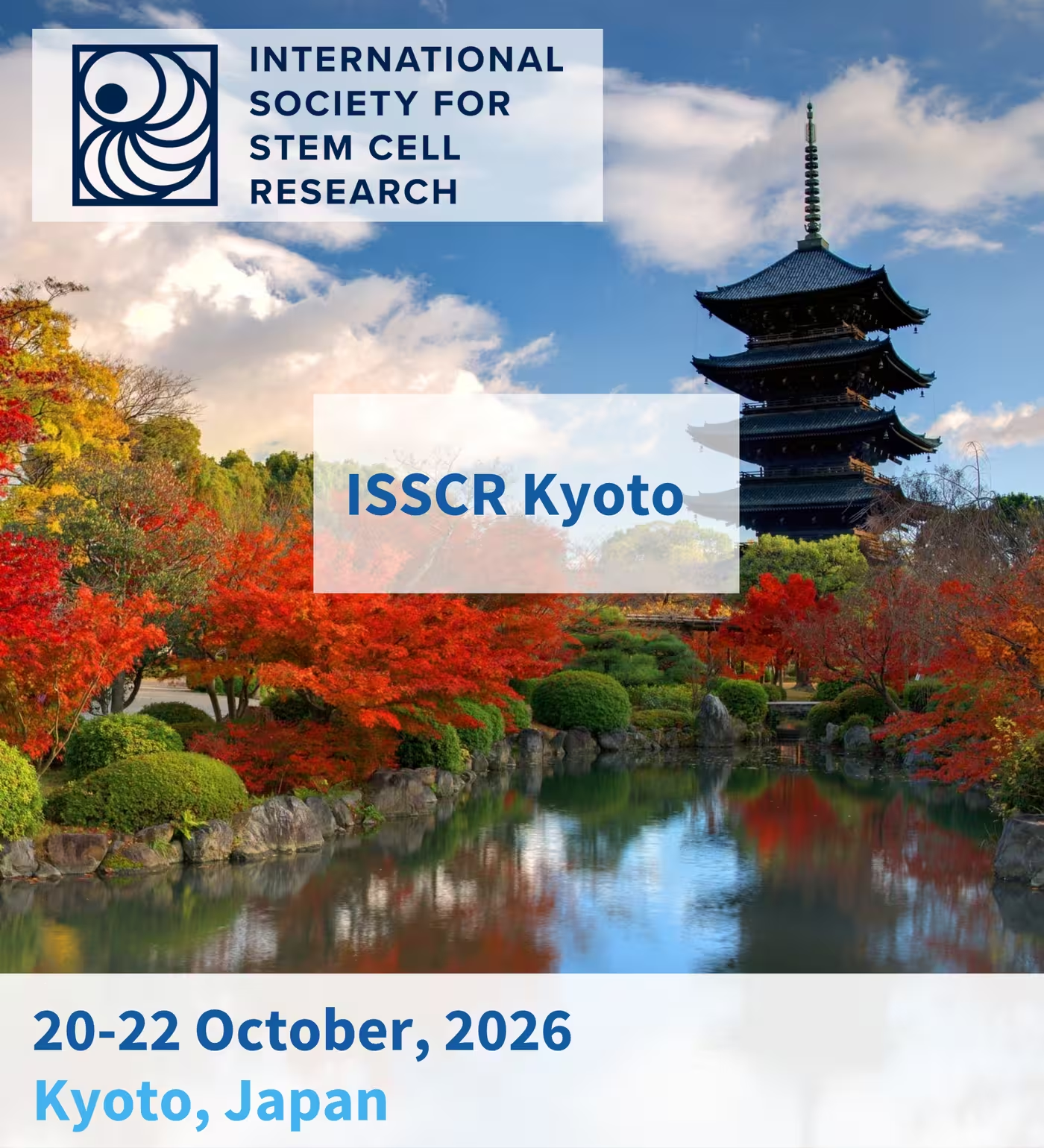
20 Years of iPSC Discovery: A Celebration and Vision for the Future

Date & Time

20-22 October, 2026

Location

Kyoto, Japan

Tags

Neuronal Cell Cultures
Organoids
Retina
Functional Phenotyping
Disease Modeling

The ISSCR 2026 Kyoto International Symposium celebrates a remarkable milestone: 20 years since the discovery of induced pluripotent stem cells (iPSCs) by Dr. Shinya Yamanaka. Bringing together the global stem cell community in the birthplace of iPSC technology, this special symposium will celebrate two decades of groundbreaking discoveries while exploring the future of the field. From fundamental stem cell biology and technological innovations to disease modeling, drug discovery, and clinical translation, the meeting offers a unique opportunity to reflect on how iPSC technology has transformed biomedical research and where the next breakthroughs may emerge. At MaxWell Biosystems, we are excited to be part of this celebration, connecting with the scientific community, exchanging ideas, and exploring how next-generation technologies can help unlock the full potential of iPSC-derived models.
Meet the MxW Team

Dr. Marie Obien

CCO | MaxWell Biosystems (Switzerland)

Dr. Urs Frey

CEO | MaxWell Biosystems (Switzerland)

Dr. Zhuoliang (Ed) Li

Product Manager, MaxOne | MaxWell Biosystems (Switzerland)

Dr. Marian Hruska-Plochan

Scientific Business Developer | MaxWell Biosystems (Switzerland)

MxW Booth

Come visit us at booth #29 to find out how MaxWell Biosystems supports stem cell research with scalable high-content functional electrophysiology, delivering high-resolution insights and quality data you can trust. Stop by to connect with the team, explore what is new, and get a glimpse of what is coming next.

Innovation Showcase

Join us for this Innovation Showcase to explore how next-generation HD-MEA technology and advanced iPSC-derived neural models are transforming functional phenotyping, disease modeling, and drug discovery. From high-resolution recordings of neuronal activity at network, single-cell, and subcellular scales to innovative approaches for modeling amyotrophic lateral sclerosis (ALS), the session will highlight how functional insights and emerging regenerative medicine strategies can advance our understanding of neurodegenerative diseases and support the development of new therapies.

October 21st, 2026 | 2:10 PM – 2:30 PM

Next-Generation Disease Modeling with Human Neural Systems: Functional Phenotyping And Therapeutic Innovation

Neurological disorders remain a major global health challenge, highlighting the urgent need for more predictive, human-relevant models and new therapeutic approaches. Induced pluripotent stem cell (iPSC)-derived neural systems, including 2D neuronal cultures and 3D brain organoids and assembloids, are transforming our ability to investigate disease mechanisms, identify functional phenotypes, and explore therapeutic strategies. Unlocking their full potential, however, requires technologies capable of capturing neuronal function across multiple scales.  

MaxWell Biosystems’ High-Density Microelectrode Array (HD-MEA) technology enables label-free, high-resolution recordings of neuronal activity, from network dynamics down to single-cell and subcellular signals. With the MaxOne and MaxTwo platforms, researchers can generate robust datasets for functional characterization, disease phenotyping, and compound screening.  

During our Innovation Showcase, Dr. Yi Sun will expand this perspective through her work on amyotrophic lateral sclerosis (ALS), including the development of isogenic “disease-in-a-dish” models and approaches for phenotypic analysis. Together, these perspectives will illustrate how advances in human disease modeling, functional phenotyping, and emerging regenerative medicine can contribute to a deeper understanding of neurodegenerative disease and support the development of new translational strategies.

‍

Dr. Marie Obien
CCO | MaxWell Biosystems (Switzerland)

Bio

Marie Obien is the Chief Commercial Officer (CCO) and a founding member of MaxWell Biosystems. Marie leads the company’s global commercial strategy, sales, business development, marketing, product management, channel partnerships, and customer success. A specialist in electrophysiology, Marie brings strong expertise in microelectrode arrays and complementary techniques, including the gold-standard patch-clamp method. She is the lead author of the most frequently cited review on MEA technology and has played a key role in shaping its adoption in neuroscience and drug discovery. Marie earned her PhD in Electrical Engineering from the Nara Institute of Science and Technology, Japan, and received neuroscience training as a postdoctoral researcher at RIKEN. She also completed the Global Innovation Program in Silicon Valley, focusing on business development and entrepreneurial leadership.

Dr. Yi Sun
Professor | Shenzhen University of Advanced Technology

Bio

Dr. Yi Eve Sun is a leading scientist in neural stem cell biology, neural repair, and autism spectrum disorders. She is a Chair Professor and Principal Investigator at the Shenzhen University of Advanced Technology (SUAT), a Foreign Full Member of the Russian Academy of Engineering (RAE), and a full member of the European Academy of Natural Sciences (EANS). She also serves as Executive Director of the Chinese National Base for International Collaboration on Stem Cells and Regenerative Medicine, and as Deputy Director of the Key Laboratory for Viral Vector Technology Research and Evaluation of Cell and Gene Therapy Products under the Chinese National Medical Products Administration. Previously, she was a tenured professor at University of California, Los Angeles (UCLA) and Associate Academic Director of its Stem Cell Institute. Her groundbreaking research includes studying MECP2 gene function in neural stem cells and establishing disease models for Rett syndrome. She was the first to use gene-editing to create non-human primate models of Rett syndrome, publishing in Cell Stem Cell and Cell. She also identified CD133-positive ependymal cells as key for neural regeneration (Cell). Her research extends to gut microbiota and immune system interactions in central nervous system (CNS) diseases and reprogramming dermal fibroblasts into rejuvenated mesenchymal stem cell-like “Prometheus Cells”, for treating ALS and aging. Dr. Sun has published over 140 papers, including in Cell, Nature, and Science, with more than 20,000 citations and an H-index of 68. She has received numerous awards, including the Alfred P. Sloan Research Fellowship, Beckman Young Investigator Award, and Shanghai Science and Technology Awards, and serves on key scientific boards.

Poster
Presentations

Poster Number: 205

Functional Profiling of hiPSC-Derived Midbrain Organoids Across Scales With High-Density Microelectrode Arrays (HD-MEAS)

Abstract

Human iPSC-derived midbrain organoids are emerging as physiologically relevant 3D models to study dopaminergic development, maturation, and dysfunction in Parkinson’s disease (PD) and related synucleinopathies. Disease-relevant phenotypes may arise as gradual changes in excitability, network dynamics, and signal propagation before overt neurodegeneration, requiring electrophysiological workflows that combine throughput, longitudinal stability, and high spatial resolution. Here, in collaboration with STEMCELL Technologies, we established a reproducible, multiscale functional profiling workflow for commercially available human iPSC-derived midbrain organoids using HD-MEAs. Organoids were plated at ~77 days onto MaxTwo HD-MEA 24-Well Plates (26'400 electrodes/well) and monitored for ~1 month. ActivityScan Assay recordings revealed progressive increases in spontaneous firing rate, spike amplitude, and active area, consistent with neuronal maturation and improved tissue-electrode coupling. Network recordings detected synchronized high frequency bursting at early timepoints that became increasingly robust and organized over maturation, with higher burst cadence and greater neuronal recruitment. Pharmacological challenge with 4-AP and TTX produced rapid, reproducible bidirectional modulation of activity, confirming assay responsiveness. Finally, AxonTracking enabled label-free mapping of somato-axon propagation at the organoid-electrode interface, providing subcellular-resolution phenotyping complementary to network-level metrics. Together, these results establish a scalable, non-invasive HD-MEA workflow for longitudinal, multiscale functional phenotyping of human iPSC-derived midbrain organoids, supporting assay development, pharmacological testing, and disease-relevant phenotyping in human 3D iPSC-derived models.

Biography

Dr. Marian Hruska-Plochan

Scientific Business Developer | MaxWell Biosystems (Switzerland)

Poster Number: 268

Unveiling hiPSC FTD Model Network nd Axonal Dysfunction With High-Density Microelectrode Arrays (HD-MEAS)

Abstract

Human iPSC-derived neuronal models provide disease-relevant platforms for studying neurodegenerative disorders such as frontotemporal dementia (FTD), while overcoming key limitations of conventional approaches. Compared with animal models, they better reflect human biology and provide scalable access to relevant cell types in a controlled setting not possible with patient CNS tissue. However, heterogeneous disorders such as FTD require robust human models combined with quantitative, longitudinal readouts across multiple biological scales. Here, in collaboration with FUJIFILM Cellular Dynamics, we describe a longitudinal workflow using high-density microelectrode arrays (HD-MEAs) to profile an iPSC-based FTD model at network, cellular, and subcellular levels. We co-cultured iCell Induced Excitatory Neurons with iCell Astrocytes 2.0 and compared an Apparently Healthy Normal (AHN) control line with an isogenic FTD model carrying a heterozygous GRN loss-of-function mutation (R493X). Using the MaxTwo 24-Well Plate HD-MEA system, with 26,400 electrodes per well, spontaneous activity was monitored from DIV 6–50. MaxLab Live ActivityScan and Network assays revealed hyperexcitability in GRN cultures, with elevated firing rates by DIV 15 (p < 0.01) and altered synchrony, including more frequent, higher-amplitude network bursts, shorter inter-burst intervals from DIV 25, and >2-fold prolonged burst durations by DIV 50 (p <0.001) relative to AHN controls. AxonTracking was used to reconstruct action potential propagation along axonal arbors. Despite increased excitability, GRN neurons showed reduced axonal branching and shorter neurites, indicating a structure–function mismatch in which hyperexcitability may partially compensate for impaired axonal function. These findings establish MaxWell HD-MEA technology as a scalable, sensitive multiscale platform for functional phenotyping of FTD-relevant neuronal dysfunction, for disease modelling and screening.

Biography

MaxWell